Key Components Of A Complete Eye Examination
Regular eye examinations are crucial for maintaining optimal eye health and detecting any underlying conditions or issues early on. A comprehensive eye examination involves a series of tests and assessments performed by an eye care professional to evaluate various aspects of vision and eye health. Understanding the key components of a complete eye examination can help individuals prioritize their eye health and ensure timely intervention when necessary.
Introduction to Eye Examinations
Eye examinations are not only about assessing visual acuity but also about evaluating the overall health of the eyes. A comprehensive eye examination typically involves several components, each serving a specific purpose in assessing different aspects of vision and eye health. From testing visual acuity to examining the internal structures of the eye, a thorough evaluation provides valuable insights into the condition of the eyes and helps identify any potential issues or abnormalities.
Visual Acuity Testing
Visual acuity testing is one of the fundamental components of an eye examination. This test measures the sharpness and clarity of vision, typically using a Snellen chart or other visual acuity charts. Patients are asked to read letters or symbols of varying sizes from a specific distance. Visual acuity testing helps identify refractive errors such as myopia (nearsightedness), hyperopia (farsightedness), and astigmatism, as well as assess the effectiveness of corrective lenses or glasses.
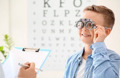
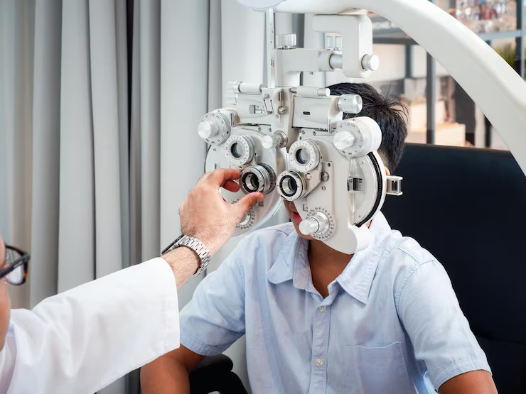
Refraction Assessment
A refraction assessment is conducted to determine the patient’s refractive error and prescribe corrective lenses if needed. The eye care professional uses a phoropter or automated refractor to measure the eye’s ability to focus light accurately on the retina. By adjusting the lenses, the examiner can determine the patient’s optimal prescription for glasses or contact lenses to correct refractive errors and improve visual clarity.
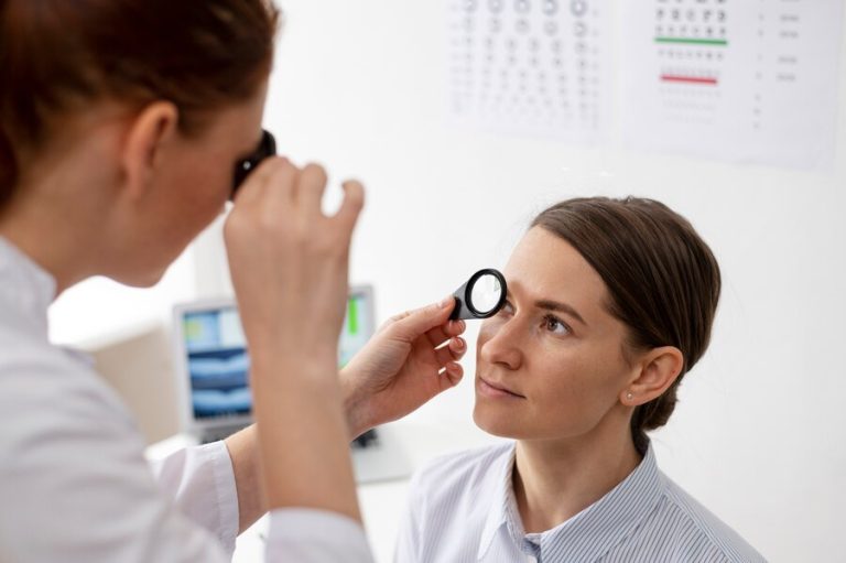
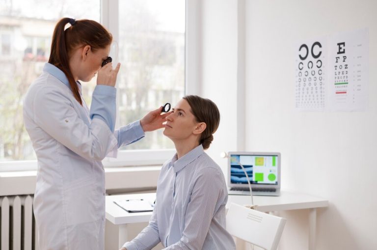
Examination of the External Eye Structures
Examination of the external eye structures involves assessing the health and integrity of the eyelids, conjunctiva, cornea, and surrounding tissues. The eye care professional examines the eyes for any signs of abnormalities, such as redness, swelling, discharge, or lesions. Additionally, the eyelid function, tear production, and blink reflex may be evaluated to identify any underlying issues affecting ocular surface health and comfort.
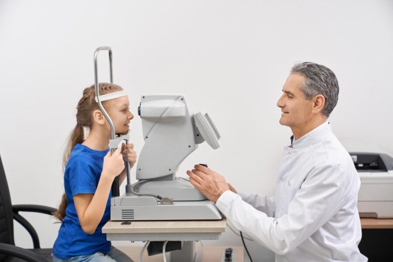
Intraocular Pressure Measurement (Tonometry)
Intraocular pressure (IOP) measurement is crucial for screening and monitoring conditions such as glaucoma, a leading cause of irreversible vision loss. Tonometry is the technique used to measure IOP, with various methods available, including application tonometry, non-contact tonometry, and tonometry with the use of specialized devices. Elevated IOP can indicate an increased risk for glaucoma and other ocular conditions, necessitating further evaluation and management.
Pupil Examination and Response Assessment
Pupil examination and response assessment evaluate the function of the pupils and the integrity of the pupillary reflexes. The eye care professional observes the size, shape, and symmetry of the pupils under different lighting conditions. Additionally, the pupillary light reflex is assessed by shining a light into each eye and observing the constriction response. Abnormalities in pupil size or reactivity may indicate neurological conditions or other underlying issues requiring further investigation.
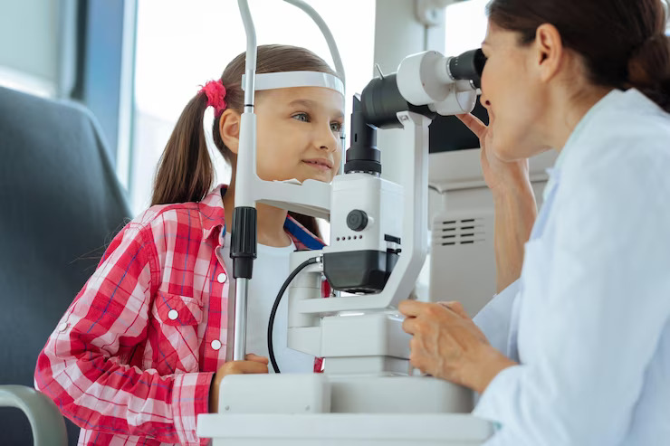
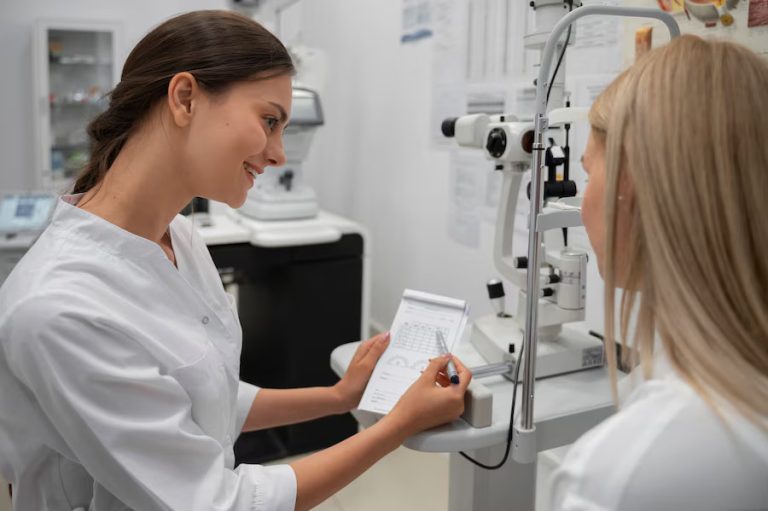
Biomicroscopy and Slit Lamp Examination
Biomicroscopy and slit lamp examination allow for detailed visualization of the anterior segment of the eye, including the cornea, iris, lens, and anterior chamber. This examination provides valuable information about the health of these structures, as well as the presence of any abnormalities or pathology. Using a slit lamp biomicroscope, the eye care professional can examine the eyes under high magnification and with various illumination techniques, enabling precise assessment and diagnosis.
Fundus Examination
Fundus examination involves evaluating the posterior segment of the eye, including the retina, optic nerve, and blood vessels, using specialized instruments such as ophthalmoscopes or fundus cameras. This examination allows for the detection of retinal disorders, such as diabetic retinopathy, macular degeneration, and retinal detachment. The health of the optic nerve and the presence of any abnormalities, such as optic nerve cupping or swelling, are also assessed during fundus examination.
Visual Field Testing
Visual field testing assesses the peripheral vision or side vision, which is essential for activities such as driving and navigating obstacles. Various methods, including confrontational visual field testing, automated perimetry, and tangent screen testing, may be used to evaluate the extent and quality of the patient’s visual field. Visual field defects can result from conditions such as glaucoma, optic nerve disorders, or neurological conditions, making this test vital for detecting and monitoring visual function abnormalities.
Conclusion
A complete eye examination encompasses various components, each playing a crucial role in assessing different aspects of vision and eye health. From testing visual acuity and refractive error to examining the external and internal structures of the eye, a thorough evaluation provides valuable insights into the condition of the eyes and helps identify any potential issues or abnormalities. By prioritizing regular eye examinations and understanding the key components involved, individuals can take proactive steps to preserve their vision and maintain optimal eye health for years to come.