Components Of A Complete Eye Examination
A comprehensive eye examination is a vital aspect of preventive healthcare, providing valuable insights into an individual’s visual health and overall well-being. This examination encompasses various components, each serving a unique purpose in assessing different aspects of ocular function and health. In order to preserve the best possible eye health, we will examine the key elements of a comprehensive eye inspection in this article, emphasizing their importance.
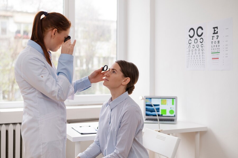
Visual Acuity Testing
Visual acuity testing is one of the fundamental components of an eye examination, aiming to assess the clarity and sharpness of vision. This test typically involves reading letters or symbols from a standardized chart at a specific distance. The results are recorded as a fraction, with 20/20 representing normal vision and numbers such as 20/40 or 20/200 indicating reduced visual acuity. Visual acuity testing helps detect refractive errors such as myopia (nearsightedness), hyperopia (farsightedness), and astigmatism, guiding the prescription of corrective lenses.
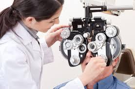
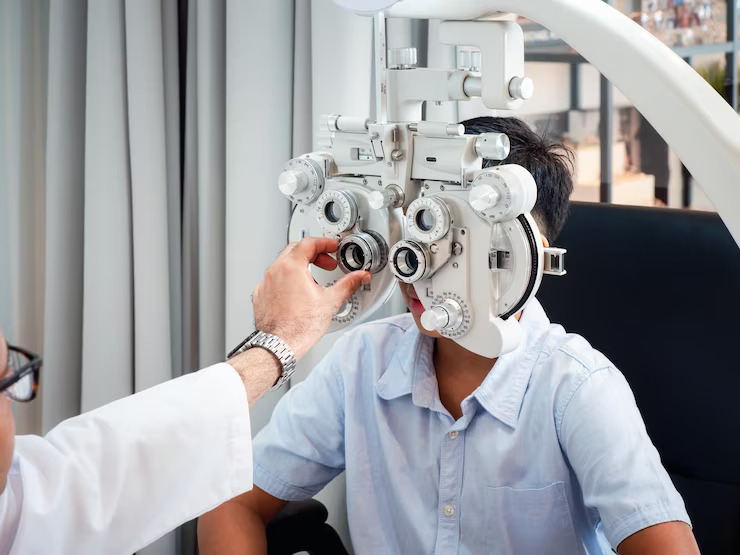
Refraction Assessment
Refraction assessment is essential for determining the appropriate prescription for glasses or contact lenses. During this component of the eye examination, the optometrist or ophthalmologist uses a phoropter or trial frame to present different lens combinations, asking the patient to provide feedback on which lenses offer the clearest vision. By refining the refractive error correction, refraction assessment aims to optimize visual acuity and alleviate symptoms of blurred vision or eyestrain.
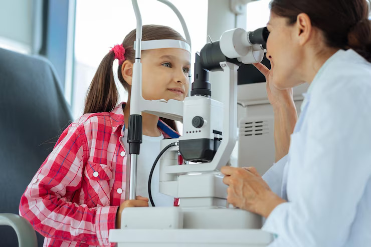
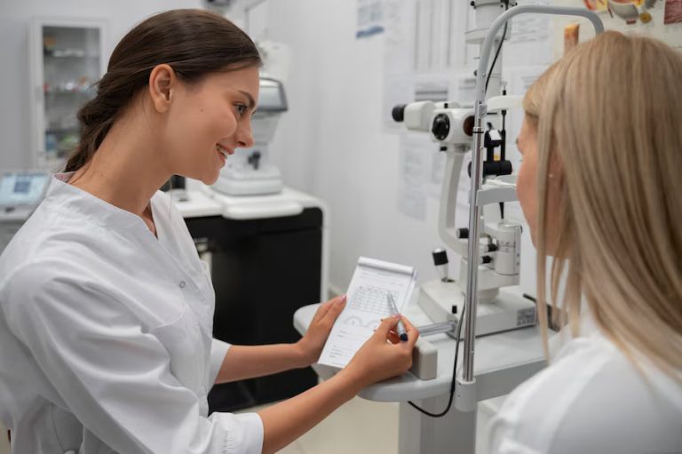
Eye Health Evaluation
The eye health evaluation component of a comprehensive eye examination involves a thorough assessment of the external and internal structures of the eye. The optometrist or ophthalmologist examines the eyelids, conjunctiva, cornea, iris, and lens for signs of abnormalities or disease. Additionally, specialized instruments such as a slit lamp biomicroscope and ophthalmoscope are used to visualize the anterior and posterior segments of the eye, including the retina, optic nerve, and macula. This examination helps detect conditions such as cataracts, glaucoma, diabetic retinopathy, macular degeneration, and retinal detachment, allowing for early intervention and treatment.
Intraocular Pressure Measurement
Intraocular pressure (IOP) measurement is crucial for assessing the risk of glaucoma, a leading cause of irreversible vision loss worldwide. Elevated IOP can damage the optic nerve and lead to progressive vision loss if left untreated. During this component of the eye examination, the optometrist or ophthalmologist uses a tonometer to measure the pressure inside the eye. Normal IOP typically ranges between 10 and 21 mmHg, although individual variations exist. Elevated IOP may prompt further evaluation and monitoring to prevent glaucoma progression.
Pupil Examination
Pupil examination is an essential component of a comprehensive eye examination, providing valuable information about neurological function and pupillary reflexes. The optometrist or ophthalmologist assesses the size, shape, symmetry, and reactivity of the pupils under different lighting conditions. Abnormalities such as anisocoria (unequal pupil size), sluggish pupillary response, or abnormal constriction may indicate underlying neurological conditions or ocular pathology, warranting further investigation.
Visual Field Testing
Visual field testing is performed to evaluate the full extent of an individual’s peripheral vision. This component of the eye examination is particularly important for detecting abnormalities such as visual field loss or defects, which may be indicative of conditions such as glaucoma, optic nerve disorders, or neurological diseases. Various techniques, including confrontation visual field testing, automated perimetry, and kinetic perimetry, may be used to assess different aspects of visual field function.
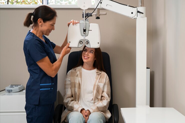
Fundus Examination
Fundus examination, also known as dilated eye examination, allows for a detailed assessment of the retina, optic nerve, and blood vessels at the back of the eye. During this component of the eye examination, the optometrist or ophthalmologist dilates the pupils using eye drops to facilitate visualization of the internal structures. A specialized instrument called an ophthalmoscope or funduscope is used to examine the retina for signs of abnormalities such as retinal detachment, macular degeneration, diabetic retinopathy, hypertensive retinopathy, or optic nerve disorders. Fundus examination plays a crucial role in the early detection, diagnosis, and management of sight-threatening conditions, enabling timely intervention to preserve vision.
Conclusion
A comprehensive eye examination encompasses a range of essential components aimed at assessing visual function, ocular health, and overall well-being. By incorporating visual acuity testing, refraction assessment, eye health evaluation, intraocular pressure measurement, pupil examination, visual field testing, and fundus examination, eye care professionals can provide a thorough assessment of an individual’s eye health status. Early detection, diagnosis, and treatment of ocular conditions through comprehensive eye examinations are essential for maintaining optimal vision and preventing vision loss. Regular eye examinations, as recommended by eye care professionals, play a critical role in preserving eye health and enhancing quality of life.
For any further queries, Plz visit drvivekgarg.in